Difference between revisions of "HaCaT Eicosanoid Analysis Results"
| Line 60: | Line 60: | ||
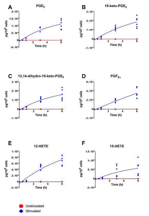
[[Image:K03_summary.jpg|none|thumb|500px|Lipid mediator levels in unstimulated (red) and stimulated with A23187 (5 µM) (blue) HaCaT keratinocytes over 6h. Where (A) PGE2, (B) 15-keto-PGE2, (C) 13,14-dihydro-15-keto-PGE2, (D) PGF2α, (E) 12-HETE and (D) 15-HETE. The data presented is from three independent experiments, each with a technical replicate. ]] | [[Image:K03_summary.jpg|none|thumb|500px|Lipid mediator levels in unstimulated (red) and stimulated with A23187 (5 µM) (blue) HaCaT keratinocytes over 6h. Where (A) PGE2, (B) 15-keto-PGE2, (C) 13,14-dihydro-15-keto-PGE2, (D) PGF2α, (E) 12-HETE and (D) 15-HETE. The data presented is from three independent experiments, each with a technical replicate. ]] | ||
| + | |||
| + | == ATP == | ||
| + | {| class="wikitable" | ||
| + | |+ style="text-align: left;" |AA-derived lipid mediators detected in the supernatant of HaCaT keratinocytes treated with ATP (2 mM), for 0.5, 1, 3 and 6h. Analysis was performed using UPLC-ESI-MS/MS. Results are expressed mean ± SD (pg/106 cells) of three independent experiments. | ||
| + | |- | ||
| + | ! rowspan="2" style="text-align: center; font-weight:bold;" | Time (h) | ||
| + | ! colspan="6" style="text-align: center; font-weight:bold;" | Mediator | ||
| + | |- | ||
| + | | style="text-align: center; font-weight:bold;" | PGE2 | ||
| + | | style="text-align: center; font-weight:bold;" | 15-keto-PGE2 | ||
| + | | style="text-align: center; font-weight:bold;" | 13,14-dihydro-15-keto-PGE2 | ||
| + | | style="text-align: center; font-weight:bold;" | PGF2α | ||
| + | | style="text-align: center; font-weight:bold;" | 12-HETE | ||
| + | | 15-HETE | ||
| + | |- | ||
| + | | style="text-align: center;" | 0 | ||
| + | | style="text-align: center;" | 1.50 ± 0.00 | ||
| + | | style="text-align: center;" | 0.37 ± 0.21 | ||
| + | | style="text-align: center;" | 2.50 ± 0.00 | ||
| + | | style="text-align: center;" | 7.50 ± 0.00 | ||
| + | | style="text-align: center;" | 1.00 ± 0.00 | ||
| + | | 2.00 ± 0.00 | ||
| + | |- | ||
| + | | style="text-align: center;" | 0.5 | ||
| + | | style="text-align: center;" | 617.77 ± 60.96 | ||
| + | | style="text-align: center;" | 1.10 ± 0.58 | ||
| + | | style="text-align: center;" | 51.51 ± 6.47 | ||
| + | | style="text-align: center;" | 96.88 ± 13.81 | ||
| + | | style="text-align: center;" | 49.56 ± 37.94 | ||
| + | | 15.82 ± 9.10 | ||
| + | |- | ||
| + | | style="text-align: center;" | 1 | ||
| + | | style="text-align: center;" | 849.12 ± 56.72 | ||
| + | | style="text-align: center;" | 3.60 ± 0.79 | ||
| + | | style="text-align: center;" | 86.88 ± 32.86 | ||
| + | | style="text-align: center;" | 148.10 ± 20.14 | ||
| + | | style="text-align: center;" | 32.28 ± 31.72 | ||
| + | | 10.95 ± 9.46 | ||
| + | |- | ||
| + | | style="text-align: center;" | 3 | ||
| + | | style="text-align: center;" | 833.49 ± 170.07 | ||
| + | | style="text-align: center;" | 2.05 ± 0.65 | ||
| + | | style="text-align: center;" | 106.43 ± 12.88 | ||
| + | | style="text-align: center;" | 170.02 ± 34.73 | ||
| + | | style="text-align: center;" | 34.05 ± 39.23 | ||
| + | | 5.70 ± 7.02 | ||
| + | |- | ||
| + | | style="text-align: center;" | 6 | ||
| + | | style="text-align: center;" | 1015.73 ± 88.65 | ||
| + | | style="text-align: center;" | 2.43 ± 1.70 | ||
| + | | style="text-align: center;" | 141.16 ± 9.62 | ||
| + | | style="text-align: center;" | 210.30 ± 43.78 | ||
| + | | style="text-align: center;" | 9.76 ± 10.52 | ||
| + | | 2.22 ± 0.54 | ||
| + | |} | ||
| + | |||
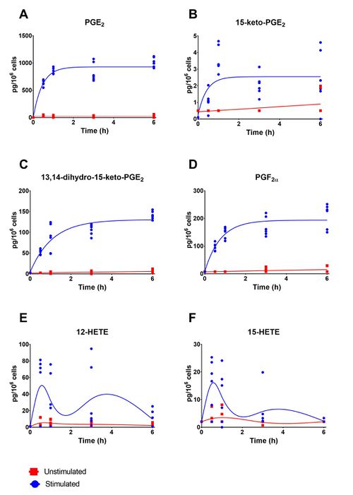
| + | [[Image:K04_eicosanoids.jpg|none|thumb|500px|Lipid mediator levels in unstimulated (red) and stimulated with ATP (2mM) (blue) HaCaT keratinocytes over 6h. Where (A) PGE2, (B) 15-keto-PGE2, (C) 13,14-dihydro-15-keto-PGE2, (D) PGF2α, (E) 12-HETE and (D) 15-HETE. The data presented is from three independent experiments, each with a technical replicate. ]] | ||
| + | |||
| + | |||
| + | == UVR == | ||
| + | |||
| + | {| class="wikitable" | ||
| + | |+ style="text-align: left;" |AA-derived lipid mediators detected in the supernatant of HaCaT keratinocytes treated with UVR (15 mJ/cm2), for 0.5, 1, 3 and 6h. Analysis was performed using UPLC-ESI-MS/MS. Results are expressed mean ± SD (pg/106 cells) of three independent experiments. | ||
| + | |- | ||
| + | ! rowspan="2" style="text-align: center; font-weight:bold;" | Time (h) | ||
| + | ! colspan="6" style="text-align: center; font-weight:bold;" | Mediator | ||
| + | |- | ||
| + | | style="text-align: center; font-weight:bold;" | PGE2 | ||
| + | | style="text-align: center; font-weight:bold;" | 15-keto-PGE2 | ||
| + | | style="text-align: center; font-weight:bold;" | 13,14-dihydro-15-keto-PGE2 | ||
| + | | style="text-align: center; font-weight:bold;" | PGF2α | ||
| + | | style="text-align: center; font-weight:bold;" | 12-HETE | ||
| + | | style="font-weight:bold;" | 15-HETE | ||
| + | |- | ||
| + | | style="text-align: center;" | 0 | ||
| + | | style="text-align: center;" | 203.29 ± 315.50 | ||
| + | | style="text-align: center;" | 0.50 ± 0.00 | ||
| + | | style="text-align: center;" | 2.00 ± 0.00 | ||
| + | | style="text-align: center;" | 0.00 ± 0.00 | ||
| + | | style="text-align: center;" | 3.50 ± 0.00 | ||
| + | | 2.30 ± 0.00 | ||
| + | |- | ||
| + | | style="text-align: center;" | 0.5 | ||
| + | | style="text-align: center;" | 1.50 ± 0.00 | ||
| + | | style="text-align: center;" | 0.75 ± 0.39 | ||
| + | | style="text-align: center;" | 2.00 ± 0.00 | ||
| + | | style="text-align: center;" | 88.72 ± 33.06 | ||
| + | | style="text-align: center;" | 135.07 ± 142.32 | ||
| + | | 40.09 ± 35.24 | ||
| + | |- | ||
| + | | style="text-align: center;" | 1 | ||
| + | | style="text-align: center;" | 69.26 ± 60.99 | ||
| + | | style="text-align: center;" | 1.05 ± 0.99 | ||
| + | | style="text-align: center;" | 2.29 ± 1.56 | ||
| + | | style="text-align: center;" | 90.56 ± 11.96 | ||
| + | | style="text-align: center;" | 131.60 ± 100.39 | ||
| + | | 46.54 ± 35.33 | ||
| + | |- | ||
| + | | style="text-align: center;" | 3 | ||
| + | | style="text-align: center;" | 1.50 ± 0.00 | ||
| + | | style="text-align: center;" | 9.61 ± 7.15 | ||
| + | | style="text-align: center;" | 3.53 ± 2.41 | ||
| + | | style="text-align: center;" | 222.44 ± 6.73 | ||
| + | | style="text-align: center;" | 103.71 ± 78.12 | ||
| + | | 58.45 ± 43.57 | ||
| + | |- | ||
| + | | style="text-align: center;" | 6 | ||
| + | | style="text-align: center;" | 5667.91 ± 4967.29 | ||
| + | | style="text-align: center;" | 152.58 ± 51.19 | ||
| + | | style="text-align: center;" | 6.21 ± 4.30 | ||
| + | | style="text-align: center;" | 1657.38 ± 297.77 | ||
| + | | style="text-align: center;" | 766.69 ± 206.41 | ||
| + | | 648.45 ± 219.84 | ||
| + | |} | ||
| + | |||
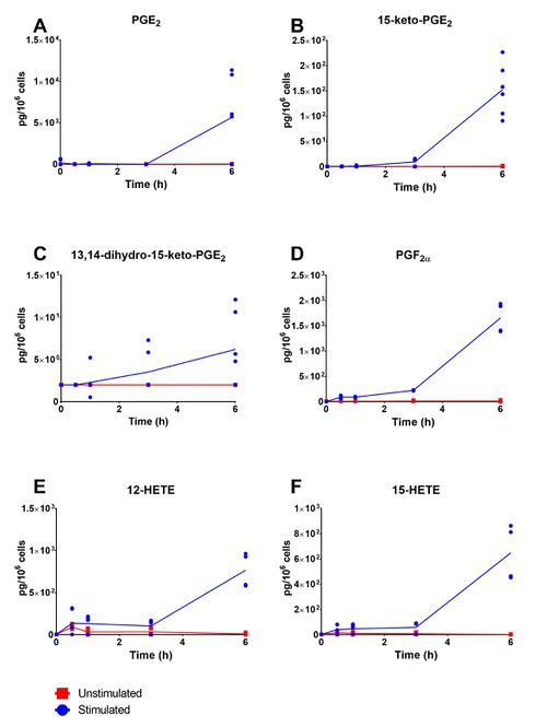
| + | [[Image:K01_eicosanoids.jpg|none|thumb|500px|Lipid mediator levels in unstimulated (red) and irradiated (15 mJ/cm2) (blue) HaCaT keratinocytes over 6h. Where (A) PGE2, (B) 15-keto-PGE2, (C) 13,14-dihydro-15-keto-PGE2, (D) PGF2α, (E) 12-HETE and (D) 15-HETE. The data presented is from three independent experiments, each with a technical replicate.]] | ||
| + | |||
| + | == Calcium ionophore + Indomethacin == | ||
| + | |||
| + | {| class="wikitable" | ||
| + | |+ style="text-align: left;" | AA-derived lipid mediators detected in the supernatant of COX inhibited HaCaT keratinocytes treated with A23187 (5 µM), for 0.5, 1, 3 and 6h. Analysis was performed using UPLC-ESI-MS/MS. Results are expressed mean ± SD (pg/106 cells) of three independent experiments. | ||
| + | |- | ||
| + | ! rowspan="2" style="text-align: center; font-weight:bold;" | Time (h) | ||
| + | ! colspan="6" style="text-align: center; font-weight:bold;" | Mediator | ||
| + | |- | ||
| + | | style="text-align: center; font-weight:bold;" | PGE2 | ||
| + | | style="text-align: center; font-weight:bold;" | 15-keto-PGE2 | ||
| + | | style="text-align: center; font-weight:bold;" | 13,14-dihydro-15-keto-PGE2 | ||
| + | | style="text-align: center; font-weight:bold;" | PGF2α | ||
| + | | style="text-align: center; font-weight:bold;" | 12-HETE | ||
| + | | 15-HETE | ||
| + | |- | ||
| + | | style="text-align: center;" | 0 | ||
| + | | style="text-align: center;" | 26.81 ± 10.56 | ||
| + | | style="text-align: center;" | 0.50 ± 0.00 | ||
| + | | style="text-align: center;" | 2.00 ± 0.00 | ||
| + | | style="text-align: center;" | 7.00 ± 0.00 | ||
| + | | style="text-align: center;" | 0.29 ± 0.00 | ||
| + | | 1.92 ± 0.94 | ||
| + | |- | ||
| + | | style="text-align: center;" | 0.5 | ||
| + | | style="text-align: center;" | 34.23 ± 22.16 | ||
| + | | style="text-align: center;" | 0.50 ± 0.00 | ||
| + | | style="text-align: center;" | 1.88 ± 0.57 | ||
| + | | style="text-align: center;" | 7.00 ± 0.00 | ||
| + | | style="text-align: center;" | 7.20 ± 10.94 | ||
| + | | 29.71 ± 60.25 | ||
| + | |- | ||
| + | | style="text-align: center;" | 1 | ||
| + | | style="text-align: center;" | 25.85 ± 9.25 | ||
| + | | style="text-align: center;" | 0.50 ± 0.00 | ||
| + | | style="text-align: center;" | 32.91 ± 28.56 | ||
| + | | style="text-align: center;" | 7.00 ± 0.00 | ||
| + | | style="text-align: center;" | 141.09 ± 24.66 | ||
| + | | 54.23 ± 51.50 | ||
| + | |- | ||
| + | | style="text-align: center;" | 3 | ||
| + | | style="text-align: center;" | 4731.85 ± 7300.78 | ||
| + | | style="text-align: center;" | 2266.13 ± 371.89 | ||
| + | | style="text-align: center;" | 1817.62 ± 907.18 | ||
| + | | style="text-align: center;" | 3268.46 ± 658.45 | ||
| + | | style="text-align: center;" | 17918.03 ± 8595.61 | ||
| + | | 55405.98 ± 17910.03 | ||
| + | |- | ||
| + | | style="text-align: center;" | 6 | ||
| + | | style="text-align: center;" | 4843.70 ± 7474.03 | ||
| + | | style="text-align: center;" | 1620.01 ± 317.98 | ||
| + | | style="text-align: center;" | 900.50 ± 199.18 | ||
| + | | style="text-align: center;" | 4754.66 ± 596.47 | ||
| + | | style="text-align: center;" | 61471.62 ± 16768.21 | ||
| + | | 105543.44 ± 41314.43 | ||
| + | |} | ||
| + | |||
| + | |||
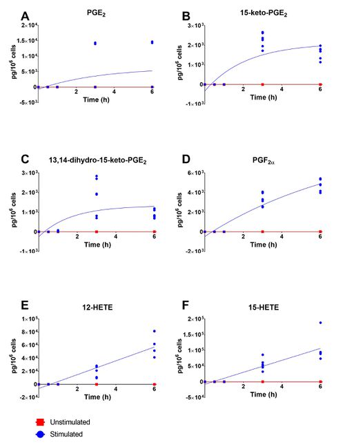
| + | [[Image:K07_eicosanoids.jpg|none|thumb|500px|Lipid mediator levels in unstimulated (red) and stimulated with A23187 (5 µM) following 1h incubation with indomethacin (10 mM) (blue) HaCaT keratinocytes, over 6h. Where (A) PGE2, (B) 15-keto-PGE2, (C) 13,14-dihydro-15-keto-PGE2, (D) PGF2α, (E) 12-HETE and (D) 15-HETE. The data presented is from three independent experiments, each with a technical replicate. ]] | ||
Revision as of 15:32, 31 May 2019
Calcium Ionophore
| Time (h) | Mediator | |||||
|---|---|---|---|---|---|---|
| PGE2 | 15-keto-PGE2 | 13,14-dihydro-15-keto-PGE2 | PGF2α | 12-HETE | 15-HETE | |
| 0 | 9.45 ± 7.56 | 0.50 ± 0.00 | 2.00 ± 0.00 | 7.00 ± 0.00 | 6.44 ± 4.40 | 3.72 ± 1.08 |
| 0.5 | 601.26 ± 162.02 | 65.52 ± 57.95 | 211.38 ± 61.05 | 106.06 ± 39.89 | 19.49 ± 10.36 | 163.26 ± 143.10 |
| 1 | 14371.34 ± 858.76 | 2136.87 ± 588.92 | 2461.84 ± 452.79 | 2787.86 ± 489.04 | 363.25 ± 96.91 | 4050.14 ± 2998.16 |
| 3 | 79576.88 ± 15355.19 | 12230.83 ± 2179.66 | 11025.93 ± 2287.76 | 19276.46 ± 3732.43 | 4872.78 ± 849.95 | 41799.69 ± 31552.30 |
| 6 | 111115.14 ± 32976.86 | 18619.32 ± 5691.99 | 15799.31 ± 5867.25 | 34965.99 ± 11250.10 | 7156.92 ± 1550.86 | 55738.98 ± 40130.05 |

Lipid mediator levels in unstimulated (red) and stimulated with A23187 (5 µM) (blue) HaCaT keratinocytes over 6h. Where (A) PGE2, (B) 15-keto-PGE2, (C) 13,14-dihydro-15-keto-PGE2, (D) PGF2α, (E) 12-HETE and (D) 15-HETE. The data presented is from three independent experiments, each with a technical replicate.
ATP
| Time (h) | Mediator | |||||
|---|---|---|---|---|---|---|
| PGE2 | 15-keto-PGE2 | 13,14-dihydro-15-keto-PGE2 | PGF2α | 12-HETE | 15-HETE | |
| 0 | 1.50 ± 0.00 | 0.37 ± 0.21 | 2.50 ± 0.00 | 7.50 ± 0.00 | 1.00 ± 0.00 | 2.00 ± 0.00 |
| 0.5 | 617.77 ± 60.96 | 1.10 ± 0.58 | 51.51 ± 6.47 | 96.88 ± 13.81 | 49.56 ± 37.94 | 15.82 ± 9.10 |
| 1 | 849.12 ± 56.72 | 3.60 ± 0.79 | 86.88 ± 32.86 | 148.10 ± 20.14 | 32.28 ± 31.72 | 10.95 ± 9.46 |
| 3 | 833.49 ± 170.07 | 2.05 ± 0.65 | 106.43 ± 12.88 | 170.02 ± 34.73 | 34.05 ± 39.23 | 5.70 ± 7.02 |
| 6 | 1015.73 ± 88.65 | 2.43 ± 1.70 | 141.16 ± 9.62 | 210.30 ± 43.78 | 9.76 ± 10.52 | 2.22 ± 0.54 |

Lipid mediator levels in unstimulated (red) and stimulated with ATP (2mM) (blue) HaCaT keratinocytes over 6h. Where (A) PGE2, (B) 15-keto-PGE2, (C) 13,14-dihydro-15-keto-PGE2, (D) PGF2α, (E) 12-HETE and (D) 15-HETE. The data presented is from three independent experiments, each with a technical replicate.
UVR
| Time (h) | Mediator | |||||
|---|---|---|---|---|---|---|
| PGE2 | 15-keto-PGE2 | 13,14-dihydro-15-keto-PGE2 | PGF2α | 12-HETE | 15-HETE | |
| 0 | 203.29 ± 315.50 | 0.50 ± 0.00 | 2.00 ± 0.00 | 0.00 ± 0.00 | 3.50 ± 0.00 | 2.30 ± 0.00 |
| 0.5 | 1.50 ± 0.00 | 0.75 ± 0.39 | 2.00 ± 0.00 | 88.72 ± 33.06 | 135.07 ± 142.32 | 40.09 ± 35.24 |
| 1 | 69.26 ± 60.99 | 1.05 ± 0.99 | 2.29 ± 1.56 | 90.56 ± 11.96 | 131.60 ± 100.39 | 46.54 ± 35.33 |
| 3 | 1.50 ± 0.00 | 9.61 ± 7.15 | 3.53 ± 2.41 | 222.44 ± 6.73 | 103.71 ± 78.12 | 58.45 ± 43.57 |
| 6 | 5667.91 ± 4967.29 | 152.58 ± 51.19 | 6.21 ± 4.30 | 1657.38 ± 297.77 | 766.69 ± 206.41 | 648.45 ± 219.84 |

Lipid mediator levels in unstimulated (red) and irradiated (15 mJ/cm2) (blue) HaCaT keratinocytes over 6h. Where (A) PGE2, (B) 15-keto-PGE2, (C) 13,14-dihydro-15-keto-PGE2, (D) PGF2α, (E) 12-HETE and (D) 15-HETE. The data presented is from three independent experiments, each with a technical replicate.
Calcium ionophore + Indomethacin
| Time (h) | Mediator | |||||
|---|---|---|---|---|---|---|
| PGE2 | 15-keto-PGE2 | 13,14-dihydro-15-keto-PGE2 | PGF2α | 12-HETE | 15-HETE | |
| 0 | 26.81 ± 10.56 | 0.50 ± 0.00 | 2.00 ± 0.00 | 7.00 ± 0.00 | 0.29 ± 0.00 | 1.92 ± 0.94 |
| 0.5 | 34.23 ± 22.16 | 0.50 ± 0.00 | 1.88 ± 0.57 | 7.00 ± 0.00 | 7.20 ± 10.94 | 29.71 ± 60.25 |
| 1 | 25.85 ± 9.25 | 0.50 ± 0.00 | 32.91 ± 28.56 | 7.00 ± 0.00 | 141.09 ± 24.66 | 54.23 ± 51.50 |
| 3 | 4731.85 ± 7300.78 | 2266.13 ± 371.89 | 1817.62 ± 907.18 | 3268.46 ± 658.45 | 17918.03 ± 8595.61 | 55405.98 ± 17910.03 |
| 6 | 4843.70 ± 7474.03 | 1620.01 ± 317.98 | 900.50 ± 199.18 | 4754.66 ± 596.47 | 61471.62 ± 16768.21 | 105543.44 ± 41314.43 |

Lipid mediator levels in unstimulated (red) and stimulated with A23187 (5 µM) following 1h incubation with indomethacin (10 mM) (blue) HaCaT keratinocytes, over 6h. Where (A) PGE2, (B) 15-keto-PGE2, (C) 13,14-dihydro-15-keto-PGE2, (D) PGF2α, (E) 12-HETE and (D) 15-HETE. The data presented is from three independent experiments, each with a technical replicate.