Difference between revisions of "Transformation of PGH2 to PGI2"
(→Parameters) |
(→Parameters) |
||
| Line 57: | Line 57: | ||
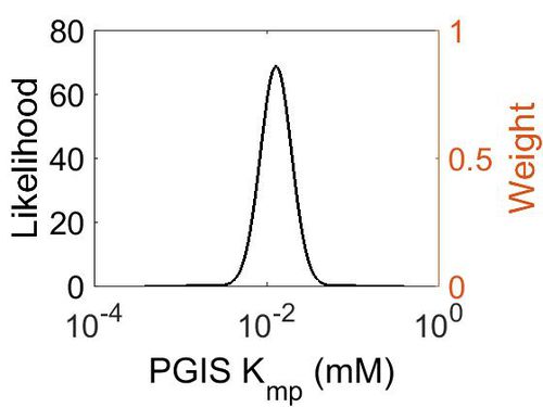
=== K<sub>mp</sub> === | === K<sub>mp</sub> === | ||
| + | {| class="wikitable" | ||
| + | |+ style="text-align: left;" | Description of the PGIS Kmp distribution | ||
| + | ! Mode (mM) !! Location parameter (µ) !! Scale parameter (σ) | ||
| + | |- | ||
| + | | 1.29E-02 || -4.182022605 || 0.414183521 | ||
| + | |} | ||
| + | |||
| + | [[Image:18.jpg|none|thumb|500px|The estimated probability distribution for PGIS Kmp. The value and weight of the literature values used to define the distribution are indicated by an orange dashed line. The x axis is plotted on a log-scale. ]] | ||
| Line 80: | Line 88: | ||
|} | |} | ||
| + | {| class="wikitable" | ||
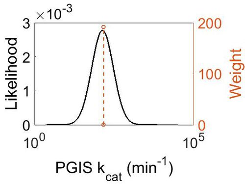
| + | |+ style="text-align: left;" | Description of the PGIS kcat distribution | ||
| + | ! Mode (min-1) !! Confidence Interval !! Location parameter (µ) !! Scale parameter (σ) | ||
| + | |- | ||
| + | | 1.41E+02 || 1.35E+00 || 5.028213628 || 0.287348692 | ||
| + | |} | ||
| − | + | [[Image:19.jpg|none|thumb|500px|The estimated probability distribution for PGIS kcat. The value and weight of the literature values used to define the distribution are indicated by an orange dashed line. The x axis is plotted on a log-scale. ]] | |
=== Enzyme concentration === | === Enzyme concentration === | ||
| Line 132: | Line 146: | ||
|} | |} | ||
| + | {| class="wikitable" | ||
| + | |+ style="text-align: left;" | Description of the PGIS concentration distribution | ||
| + | ! Mode (mM) !! Confidence Interval !! Location parameter (µ) !! Scale parameter (σ) | ||
| + | |- | ||
| + | | 1.13E+02 || 4.13E+00 || 5.67E+00 || 9.68E-01 | ||
| + | |- | ||
| + | | | ||
| + | |} | ||
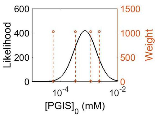
| + | [[Image:164.jpg|none|thumb|500px|The estimated probability distribution for PGIS concentration. The value and weight of the literature values used to define the distribution are indicated by an orange dashed line. The x axis is plotted on a log-scale. ]] | ||
=== K<sub>eq</sub> === | === K<sub>eq</sub> === | ||
| Line 156: | Line 179: | ||
|- | |- | ||
|} | |} | ||
| + | |||
| + | {| class="wikitable" | ||
| + | |+ style="text-align: left;" | Description of the PGIS Keq distribution | ||
| + | ! Mode !! Confidence Interval !! Location parameter (µ) !! Scale parameter (σ) | ||
| + | |- | ||
| + | | 9.35E-01 || 1.00E+01 || 7.30E-01 || 8.90E-01 | ||
| + | |} | ||
| + | |||
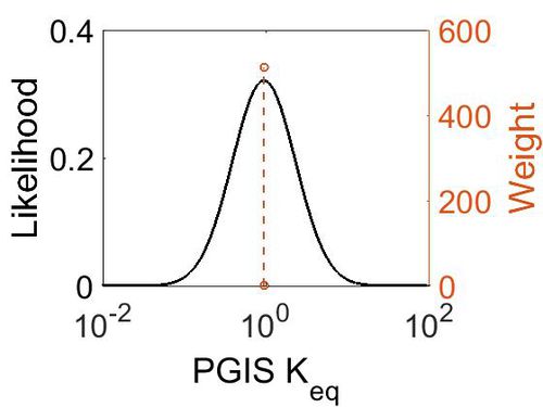
| + | [[Image:20.jpg|none|thumb|500px|The estimated probability distribution for PGIS kcat. The value and weight of the literature values used to define the distribution are indicated by an orange dashed line. The x axis is plotted on a log-scale. ]] | ||
== References == | == References == | ||
Revision as of 07:16, 15 May 2019
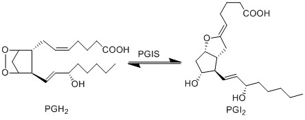
PGH2 is metabolised into the prostacyclin PGI2, by PGIS. This species has been associated with influencing the permeability of vascular compartment (Murata, Ushikubi et al. 1997). Similarly to the TX species, prostacyclins are only found in low concentration and are associated with infiltrating cells from the vascular compartment (Sugimoto, Arai et al. 2006).
Contents
Reaction
Chemical equation

Rate equation
Parameters
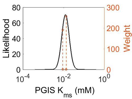
Kms
| Value | Units | Species | Notes | Reference |
|---|---|---|---|---|
| 1.33E-02 ± 1.40E-03 | 
|
Human | Expression Vector: Bovine Endothelial and Aorta Cells
Enzyme: Human PGIS pH:7.4 Temperature: 23 |
[1] |
| 9.00E-03 ± 5.00E-03 | 
|
Bovine | Expression Vector: Bovine Endothelial and Aorta Cells
Enzyme: Bovine PGIS pH:7.4 Temperature: 24 |
[2] |
| Mode (mM) | Confidence Interval | Location parameter (µ) | Scale parameter (σ) |
|---|---|---|---|
| 1.24E-02 | 2.46E+00 | -4.22E+00 | 4.20E-01 |
Kmp
| Mode (mM) | Location parameter (µ) | Scale parameter (σ) |
|---|---|---|
| 1.29E-02 | -4.182022605 | 0.414183521 |
kcat
| Value | Units | Species | Notes | Reference |
|---|---|---|---|---|
| 147 ± 45 | per minute | Cattle | Expression Vector: E. Coli
Enzyme: Bovine PGIS pH:7.4 Temperature: 24 |
[2] |
| Mode (min-1) | Confidence Interval | Location parameter (µ) | Scale parameter (σ) |
|---|---|---|---|
| 1.41E+02 | 1.35E+00 | 5.028213628 | 0.287348692 |
Enzyme concentration
| Value | Units | Species | Notes | Reference |
|---|---|---|---|---|
| 412 | 
|
Human | Expression Vector: Urinary bladder
Enzyme: PGIS pH: 7.5 Temperature: 37 °C |
[3] |
| 206 | 
|
Human | Expression Vector: Lung
Enzyme: PGIS pH: 7.5 Temperature: 37 °C |
[3] |
| 60.1 | 
|
Human | Expression Vector: Esophagus
Enzyme: PGIS pH: 7.5 Temperature: 37 °C |
[3] |
| 9.93 | 
|
Human | Expression Vector: Oral Cavity
Enzyme: PGIS pH: 7.5 Temperature: 37 °C |
[4] |
| Mode (mM) | Confidence Interval | Location parameter (µ) | Scale parameter (σ) |
|---|---|---|---|
| 1.13E+02 | 4.13E+00 | 5.67E+00 | 9.68E-01 |
Keq
| Gibbs Free Energy Change | Units | Species | Notes | Reference |
|---|---|---|---|---|
| 0.04 | kcal/mol | Not stated | Estimated
Enzyme: Transacylase Substrate: Product: pH: 7.3 ionic strength: 0.25 |
[5] |
| Mode | Confidence Interval | Location parameter (µ) | Scale parameter (σ) |
|---|---|---|---|
| 9.35E-01 | 1.00E+01 | 7.30E-01 | 8.90E-01 |
References
- ↑ H. C. Yeh, P. Y. Hsu, Characterization of heme environment and mechanism of peroxide bond cleavage in human prostacyclin synthase. Biochim Biophys Acta. 2005 Dec 30;1738(1-3):121-32.
- ↑ 2.0 2.1 Hara S. , Isolation and molecular cloning of prostacyclin synthase from bovine endothelial cells. J Biol Chem. 1994 Aug 5;269(31):19897-903. Cite error: Invalid
<ref>tag; name "Hara1994" defined multiple times with different content - ↑ 3.0 3.1 3.2 M. Kim A draft map of the human proteome Nature, 2014 509, 575–581
- ↑ M. Wilhelm Mass-spectrometry-based draft of the human proteome Nature, 2014 509, 582–587
- ↑ Caspi et al 2014, "The MetaCyc database of metabolic pathways and enzymes and the BioCyc collection of Pathway/Genome Databases," Nucleic Acids Research 42:D459-D471
